Confocal Laser Scanning Microscopy (CLSM)
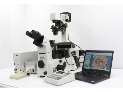
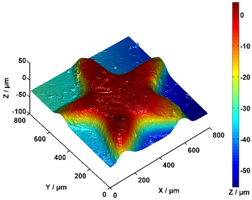
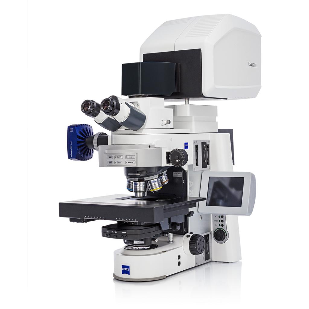
Confocal Laser Scanning Microscopy (CLSM) is an advanced optical imaging technique that uses point illumination and spatial filtering (a pinhole) to capture precise, high-contrast images of fluorescently labeled or naturally fluorescent samples. By scanning the sample point-by-point and layer-by-layer, CLSM produces optically sectioned images that can be reconstructed into 3D visualizations of cells, tissues, biomaterials, and surfaces.
Because CLSM eliminates out-of-focus light, it offers superior contrast compared to widefield fluorescence microscopy and is ideal for studying biofilms, engineered tissues, hydrogels, polymer surfaces, coatings, and biological interfaces in high detail.
Use of Confocal Laser Scanning Microscopy
CLSM is used to analyze fluorescently labeled samples and materials with natural autofluorescence.
It allows researchers to:
• Visualize 3D structures, surface morphology, and spatial organization
• Assess material–cell interactions and biological integration
• Study biofilm formation, viability, and architectural heterogeneity
• Evaluate polymer blends, coatings, and microstructures using fluorescent probes
• Track penetration of dyes, drugs, nanoparticles, and antimicrobial agents
• Image hydrated, soft, or delicate samples without physical sectioning
• Quantify fluorescence intensity, distribution, and spatial co-localization
Applications of CLSM
CLSM supports a wide range of biomedical, materials science, and industrial research applications:
• Biomaterials & Tissue Engineering: 3D visualization of scaffolds, hydrogel networks, and cellular infiltration
• Biofilms & Microbiology: architecture, depth, viability mapping, live/dead staining
• Medical Devices: coating uniformity, interface analysis, polymer–tissue interaction
• Polymers & Coatings: fluorescent dye penetration, crosslink uniformity, microdefect identification
• Nanomaterials: tracking particles inside tissue models, gels, or polymer matrices
• Cell Biology: localization of proteins, cytoskeletal structures, and intracellular transport
Sample Analysis Process
1. Sample Submission & Objective Review
- Client provides sample details, desired fluorophores, and imaging goals
• We evaluate preparation needs (staining, mounting, hydration)
2. Sample Preparation
Depending on sample type:
• Fluorescent labeling using live/dead, DAPI, FITC, or custom stains
• Mounting in imaging chambers or fluid wells
• Hydrated or biological samples kept under physiological conditions
• For polymers/coatings, fluorescent contrast agents may be applied
3. CLSM Imaging & Optical Sectioning
We use depth-resolved imaging modes such as:
• Z-stack scanning: layer-by-layer imaging for 3D reconstructions
• Multi-channel fluorescence: simultaneous imaging of multiple stains
• Reflection/confocal mode: for non-fluorescent surfaces and thin films
• Live imaging: time-lapse acquisition for dynamic studies
• 3D rendering & volumetric analysis
4. Data Processing & Reporting
- High-resolution 2D and 3D images with scale bars
• Fluorescence intensity quantification
• Depth measurements and viability ratios (for biofilms/cells)
• Annotated report aligned with study objectives
Why Choose Materials Metric for Your CLSM Analysis
Materials Metric provides precise, high-contrast optical sectioning and fluorescence imaging for complex biological and material systems.
Our CLSM analysis enables in-depth visualization of microscale and nanoscale interactions that cannot be captured by conventional microscopy.
Our CLSM services include:
- 3D imaging of hydrogels, tissues, biomaterials, and polymer structures
• Live/dead staining and viability analysis for biofilms and cell populations
• Multi-channel fluorescence imaging for co-localization studies
• Surface and interface evaluation of coatings and engineered materials
• Quantitative measurement of thickness, penetration depth, fluorescence intensity
• Specialized handling of hydrated, soft, or biological samples
• Detailed, annotated reports customized to your R&D or validation needs
By integrating CLSM findings with SEM, AFM, biochemical assays, and mechanical testing, Materials Metric provides a complete understanding of material–biological interactions and microscale structural performance.