TL;DR:
- Choosing the correct assay type is crucial for reliable drug discovery and regulatory approval.
- Validation following ICH Q2(R2) ensures assay accuracy, precision, and specificity used in pharma.
- Troubleshooting common interference helps prevent false positives and costly late-stage failures.
Not all biological assays are created equal, and in pharmaceutical drug discovery, that distinction matters enormously. Choosing the wrong assay format can derail a promising lead series, generate misleading data, or fail regulatory scrutiny at the worst possible moment. Assays in biology are laboratory procedures designed to measure the activity, concentration, or function of a biological molecule or process, and they sit at the center of every high-throughput screening campaign and quality assurance protocol your team runs. This guide covers assay categories, development workflows, validation requirements, and the troubleshooting strategies that separate reliable data from costly artifacts.
Table of Contents
- What is an assay in biology?
- Types of biological assays: Biochemical vs. cell-based
- Assay development workflows and optimization
- Assay validation: ICH Q2(R2) guidelines and QA essentials
- Common assay pitfalls and advanced troubleshooting
- Expert perspective: The evolving role of assays in modern drug development
- Connect advanced assay strategies with your lab’s success
- Frequently asked questions
Key Takeaways
| Point | Details |
|---|---|
| Assays underpin drug development | Biological assays are critical for screening new drug candidates and ensuring pharmaceutical quality. |
| Choose the right assay type | Select between biochemical and cell-based assays depending on throughput needs and physiological relevance. |
| Follow ICH validation standards | Complying with ICH Q2(R2) guidelines confirms assay accuracy and reliability. |
| Mitigate common pitfalls | Apply controls and counterscreens to avoid interference and artifacts in assay data. |
| Stay adaptive with innovation | Continuous validation and adopting advanced analytics support robust, forward-looking assay strategies. |
What is an assay in biology?
At its core, a biological assay is a method to measure the activity, concentration, or function of a molecule, cell, or biological process in a controlled laboratory environment. That definition sounds simple, but the implications are vast. Every decision your team makes, from target selection and lead identification to final product release, rests on assay data.
In pharmaceutical development, assays serve two distinct but equally critical purposes. First, they power drug discovery, enabling high-throughput screening (HTS) of compound libraries against validated biological targets. Second, they form the backbone of quality assurance, providing the quantitative evidence needed to confirm that every batch meets specifications before it reaches patients. Neglecting either role creates risk.
The two broad categories you will encounter most often are:
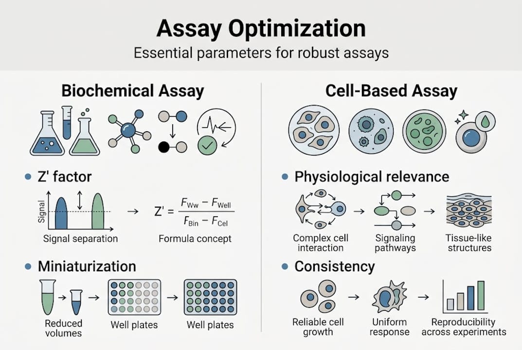
- Biochemical assays: Measure direct molecular interactions such as enzyme kinetics, receptor binding affinity, and substrate turnover. These are highly controlled and reproducible.
- Cell-based assays: Evaluate biological responses within living cells, capturing pathway activation, phenotypic changes, and downstream signaling effects that biochemical formats simply cannot replicate.
Both categories support the full arc of product development, but selecting the right one at the right stage is what separates efficient pipelines from ones plagued by late-stage failures.
“The assay is the experiment’s anchor. Get it wrong, and every downstream decision is built on sand.”
Your team should also understand that testing assay methods must align with the intended use from day one. An assay optimized for rapid screening may not be directly transferable to a regulated QA environment without significant re-validation.
Types of biological assays: Biochemical vs. cell-based
Understanding the fundamental differences between assay types is not academic, it directly shapes screening strategy and resource allocation. Biochemical assays measure enzyme activity and binding, while cell-based assays capture phenotypic and pathway-level responses. Each has a defined role in a tiered drug discovery program.
Biochemical assays excel in primary HTS because they offer low variability, easy miniaturization, and consistent Z’ factors at or above 0.5, the benchmark for HTS-ready assay quality. They measure isolated molecular events, which makes them fast and cost-effective for screening hundreds of thousands of compounds.
Cell-based assays provide physiological relevance that no biochemical format can match. When you need to know whether a compound actually modulates a pathway in a living cell, or whether it triggers toxicity, cell-based formats deliver that answer. The tradeoff is greater complexity, higher cost per well, and more variability that demands tighter experimental controls.

| Parameter | Biochemical assay | Cell-based assay |
|---|---|---|
| Throughput | Very high | Moderate to high |
| Reproducibility | High (Z’ ≥ 0.5 to 1.0) | Moderate |
| Physiological relevance | Low to moderate | High |
| Cost per well | Low | Moderate to high |
| Automation suitability | Excellent | Good |
| Complexity | Low | High |
A well-designed drug discovery program uses both in sequence. Biochemical assays handle the initial screen, and cell-based assays confirm activity in a more biologically meaningful context. Reviewing assay types and validation criteria before committing to a format prevents costly late-stage pivots.
Pro Tip: If your cell-based assay Z’ falls below 0.5 during development, do not proceed to screening. Investigate cell passage number, seeding density, and compound solubility before scaling up.
For teams working across multiple molecule classes, exploring molecular assays in pharma provides additional format guidance tailored to nucleic acid and protein targets.
Assay development workflows and optimization
Building a robust assay from concept to an HTS-ready campaign is a structured process, and shortcuts at any stage tend to resurface as expensive failures downstream. A sound development workflow moves through target biology, format selection, reagent optimization, and validation before screening begins.
Here is the standard stepwise process your team should follow:
- Target biology assessment: Confirm the target is tractable and the biological question is clearly defined.
- Detection format selection: Choose the signal modality (fluorescence, luminescence, absorbance, label-free) based on throughput needs and compound interference risk.
- Reagent sourcing and characterization: Validate all biological reagents, including enzymes, antibodies, and cell lines, before use.
- Assay miniaturization: Transition from large-volume development formats to 384-well or 1536-well plates to reduce reagent costs and increase throughput.
- Automation integration: Confirm that liquid handling, plate reading, and data capture systems are compatible with the chosen format.
- Statistical validation: Confirm Z’ factor, signal-to-background ratio, and coefficient of variation across multiple days and operators.
Optimization is iterative. The table below summarizes the key parameters your team should track:
| Optimization parameter | Target value | Why it matters |
|---|---|---|
| Z’ factor | ≥ 0.5 | Confirms assay window is adequate for HTS |
| Signal-to-background ratio | ≥ 3 | Separates real signal from noise |
| Coefficient of variation (CV) | ≤ 10% | Ensures run-to-run reproducibility |
| Compound interference rate | Minimize | Reduces false positive burden |
For teams managing rapid timelines, rapid screening assays and molecular assay development resources can accelerate format decisions.
Pro Tip: Monitor your Z’ factor throughout optimization, not just at the end. A Z’ that drifts below 0.5 mid-campaign often flags hidden reagent stability issues or interference from the DMSO vehicle, both of which are correctable if caught early.
Assay validation: ICH Q2(R2) guidelines and QA essentials
Validation is not optional. It is the regulatory and scientific foundation that confirms your assay is fit for its intended purpose. For pharmaceutical applications, ICH Q2(R2) defines the core parameters your team must demonstrate: accuracy (98 to 102% recovery), precision (RSD ≤ 2%), specificity, linearity (r² ≥ 0.99), range (80 to 120%), and both limit of detection and limit of quantitation.
Each parameter answers a specific scientific question about your method’s performance. Accuracy confirms that your assay returns the true value. Precision confirms it does so consistently. Specificity confirms it is not fooled by matrix effects or structurally related compounds. Linearity confirms the signal responds proportionally across the relevant concentration range.
Key validation steps your team must complete include:
- Accuracy studies: Spike known amounts into matrix and calculate percent recovery across at least three concentration levels.
- Precision assessment: Run intra-day and inter-day replicates to characterize repeatability and intermediate precision.
- Specificity testing: Challenge the assay with potential interferents, degradation products, and excipients.
- Linearity determination: Prepare calibration standards across the full working range and confirm r² ≥ 0.99.
- Robustness evaluation: Deliberately vary parameters like pH, temperature, and reagent lot to find the assay’s tolerance limits.
- LOD/LOQ determination: Establish the lowest detectable and quantifiable signal with acceptable accuracy.
Statistical callout: ICH Q2(R2) requires a minimum of nine determination points across three concentration levels for linearity, and HPLC methods must demonstrate a resolution factor Rs ≥ 2.0 to confirm peak separation.
For pharmaceutical QA, forced degradation studies are particularly important. They expose the assay to stress conditions, heat, light, hydrolysis, oxidation, to confirm it can distinguish intact drug substance from degradation products. Confirming assay validation and regulation alignment early keeps your program on track. Teams also benefit from integrating batch consistency in validation protocols to catch lot-to-lot variability before it reaches the clinic.
Common assay pitfalls and advanced troubleshooting
Even well-designed assays encounter interference. Recognizing the source quickly prevents wasted screening cycles and misleading SAR data. The most frequent culprits are fluorescence interference, compound aggregation, and redox cycling artifacts, all of which can generate false positives that look compelling until orthogonal confirmation exposes them.
Common sources of assay interference include:
- Autofluorescence: Highly conjugated compounds emit signal in the same channel as your probe, inflating apparent activity.
- Compound aggregation: At higher concentrations, many compounds form colloidal aggregates that non-specifically inhibit proteins.
- PAINS (pan-assay interference compounds): Reactive chemical scaffolds that interfere across multiple assay formats, generating systematic false positives.
- Redox cycling: Certain quinones and nitroaromatics consume NADH or generate reactive oxygen species, distorting enzyme activity readouts.
- Luciferase inhibition: Compounds that directly inhibit the reporter enzyme create false hits in luciferase-based cell viability assays.
“The most dangerous interference is the kind you do not know you have. Counterscreens are not optional; they are the scientific controls that make your primary data trustworthy.”
A structured troubleshooting workflow for a compromised assay should include:
- Add 0.01% Triton X-100 or equivalent detergent to break up aggregator compounds and retest.
- Run compound-only wells (no enzyme or cells) to quantify intrinsic signal from the compound itself.
- Deploy an orthogonal assay using a different detection chemistry to confirm or refute the primary hit.
- Filter compound libraries against known common assay interference databases before screening.
- Retest confirmed hits at multiple concentrations using a direct-binding or label-free format.
Addressing interference systematically at the development stage, rather than the hit confirmation stage, saves significant time and resources across the full discovery campaign.
Expert perspective: The evolving role of assays in modern drug development
There is a tempting narrative in biopharma right now: that AI, machine learning, and real-time analytics will make traditional assay development obsolete. We disagree, and the data supports our position. PAT and RTRT technologies integrated with AI/ML prediction must still be grounded in validated, robust biological data to satisfy ICH lifecycle requirements. Digital tools amplify good assay science; they do not replace it.
What we are seeing is a productive convergence. Direct-detection formats, label-free technologies like surface plasmon resonance and bio-layer interferometry, and proximity-based assays such as AlphaScreen are gaining traction because they reduce interference risk and improve data quality. These are not replacements for biochemical and cell-based approaches but additions to a tiered toolkit.
The teams that will lead in drug discovery are those that pair rigorous traditional validation with selective adoption of new formats, guided by the specific biological question at hand. Regularly revisiting older assays with updated validation protocols is equally important. As platforms evolve and molecule classes shift, old assays may carry new risks that a fresh validation cycle would catch. We encourage your team to treat validation as a living process, not a one-time milestone. Staying current with molecular assay trends is one practical way to keep that perspective active.
Connect advanced assay strategies with your lab’s success
Building and validating high-performance biological assays requires more than protocol expertise. It demands precise analytical characterization of every reagent, substrate, and formulation component supporting your assay system.

At Materials Metric, we function as an extension of your research and QA team, providing specialized analytical testing methods and material characterization techniques that strengthen assay reliability from the reagent level up. Whether your team needs chemical verification of assay components, surface characterization of microplate materials, or chemical and microscopy CRO services to support method troubleshooting, we have the instrumentation and expertise to deliver results aligned with GLP/GMP and ISO 9001:2015 standards. Contact us to discuss how we can support your next assay development or validation project.
Frequently asked questions
What is the Z’ factor and why is it important in assays?
The Z’ factor measures assay robustness by quantifying the separation between positive and negative controls. Values at or above 0.5 confirm the assay is reliable and ready for high-throughput screening campaigns.
How do I avoid common assay interference such as PAINS or autofluorescence?
Using counterscreens and orthogonal assays alongside detergent controls and compound-only wells allows your team to detect and reduce interference before it contaminates your hit list.
What are the main differences between biochemical and cell-based assays?
Biochemical assays are highly reproducible and suited for primary HTS, while cell-based assays offer physiological relevance by capturing pathway and phenotypic responses in living cells, making them ideal for secondary confirmation.
Why is ICH Q2(R2) validation required for pharmaceutical assays?
ICH Q2(R2) establishes that your method delivers accurate, reproducible, and specific results across the full working range, providing the regulatory evidence needed for product quality decisions and submissions.