Advanced Microscopy Techniques
Advanced microscopy is essential for understanding material structure, surface behavior, and biological interactions across length scales. High-resolution imaging enables precise visualization of morphology, defects, interfaces, and spatial organization that directly influence performance, safety, and reliability. At Materials Metric, we apply advanced microscopy techniques to interrogate materials and biological systems from the macro to the nanoscale. Our imaging workflows support polymers, metals, ceramics, composites, biomaterials, tissues, implants, and hybrid constructs, delivering clear, reproducible insights into surface topology, internal microstructure, and material–biological interfaces for research, product development, failure analysis, and regulatory support.
Microscopy lies at the core of our materials evaluation capabilities. We employ a range of imaging techniques to visualize surface morphology, internal microstructure, and nanoscale features with unmatched precision.
Techniques Include:
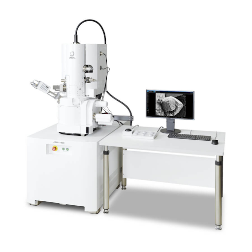
- Scanning Electron Microscopy (SEM):
Provides high-resolution imaging of surface morphology, texture, and microstructural features. Using a focused electron beam, SEM reveals nanoscale details with exceptional depth of field and enables elemental mapping through Energy Dispersive X-ray Spectroscopy (EDS). Ideal for studying coatings, fractures, and micro-defects in polymers, metals, and composites.
- Atomic Force Microscopy (AFM):
Captures ultra-high-resolution 3D topographical maps by measuring atomic-level interactions between a cantilever probe and the sample surface. AFM quantifies surface roughness, adhesion, stiffness, and elasticity, making it essential for nanomaterials, thin films, and biological samples that are difficult to analyze using electron microscopy.
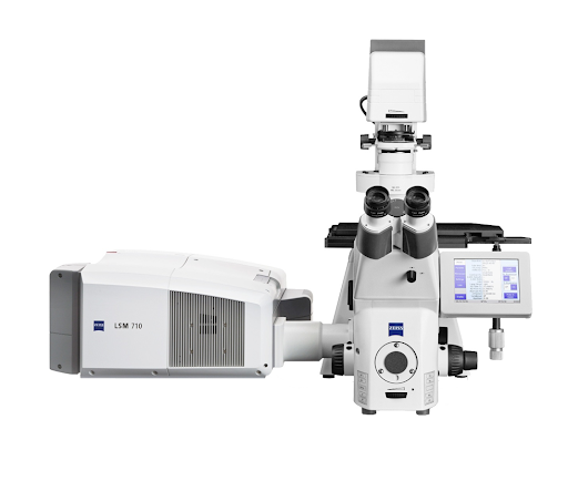
- Confocal Laser Scanning Microscopy (CLSM):
Uses point illumination and optical sectioning to create high-resolution 3D images of thick or heterogeneous materials. Confocal microscopy provides detailed visualization of biological tissues, polymers, and coated surfaces, enabling depth profiling and fluorescence overlay for structural interpretation.
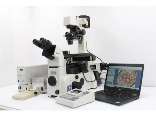
- Fluorescence Microscopy:
Employs fluorescent dyes or markers to visualize and quantify biological or material-specific features. It enables selective imaging of cells, biofilms, and functionalized biomaterials, providing insight into viability, molecular localization, and chemical labeling in both static and dynamic systems.
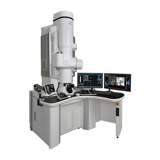
- Transmission Electron Microscopy (TEM):
Delivers atomic-scale imaging and crystallographic analysis by transmitting an electron beam through ultrathin specimens. TEM reveals internal structure, defects, and phase boundaries with unparalleled spatial resolution and supports electron diffraction studies for crystal orientation and lattice structure analysis.

