Fluorescence Microscopy
Fluorescence Microscopy is an optical imaging technique that uses fluorescent dyes, probes, or naturally fluorescent molecules to visualize specific structures, proteins, microorganisms, or material components. By exciting fluorophores with specific wavelengths of light, the microscope captures emitted light to generate high-contrast images highlighting only the stained or fluorescent regions of interest.
Fluorescence microscopy is widely used to evaluate biological samples, biofilms, tissues, hydrogels, nanoparticles, coatings, and engineered biomaterials due to its sensitivity and selectivity. It can also be combined with advanced staining techniques for viability analysis, localization, and molecular tracking.
Use of Fluorescence Microscopy
Fluorescence microscopy is used to visualize and quantify fluorescently labeled components in biological and material samples. It enables researchers to:
- Detect specific molecules, cells, or microstructures using fluorescent tags
- Evaluate cell attachment, viability, and distribution on biomaterials
- Characterize biofilm architecture and live/dead populations
- Track penetration of dyes, drugs, nanoparticles, or antimicrobial agents
- Assess polymer blends or coatings using fluorescent contrast agents
- Observe interactions between cells, tissues, and engineered materials
- Perform multi-channel imaging for protein localization and co-expression
Applications of Fluorescence Microscopy
Fluorescence microscopy is widely used in biomedical and materials research:
- Biofilms & Microbiology: visualization of bacterial communities, viability mapping
- Biomaterials & Tissue Engineering: cell infiltration, scaffold characterization, hydrogel uniformity
- Medical Devices: coating integrity, surface-biology interaction
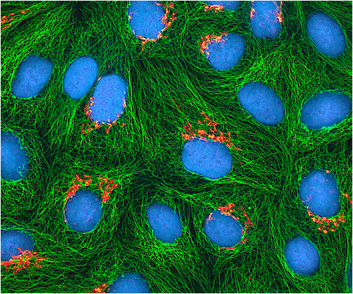
- Cell Biology: protein localization, cytoskeletal imaging, intracellular staining
- Nanomaterials: uptake or surface localization of particles
- Pharmaceutical Research: drug penetration, delivery tracking
Sample Analysis Process
1. Sample Submission & Objective Review
- Provide sample type, fluorophore(s), and imaging goals
- We determine appropriate filters, channels, and staining requirements
2. Sample Preparation
- Live/dead staining for cells or biofilms
- DAPI, FITC, TRITC, or custom fluorophore labeling
- Mounting in imaging chambers or hydrated environments
- For non-biological materials, fluorescent dyes may be added for contrast
3. Imaging Acquisition
- Multi-channel fluorescence imaging
- Z-stack acquisition for thicker samples
- Time-lapse imaging if dynamic processes are being monitored
- Brightfield or phase-contrast imaging as complementary channels
4. Data Processing & Reporting
- Fluorescence intensity maps
- Cell count, viability ratios, and distribution analysis
- Depth profiles and reconstructed 3D views
- Fully annotated report summarizing findings
Why Choose Materials Metric for Your Fluorescence Microscopy Analysis
Materials Metric provides sensitive, high-contrast fluorescence imaging ideal for evaluating biological responses, material interactions, and functional labeling.
Our capabilities include:
- Multi-channel fluorescence and viability imaging
- 2D and 3D visualization of cells, biofilms, tissues, and biomaterials
- Quantification of fluorescence intensity, depth, and distribution
- Specialized sample handling for hydrated, soft, or delicate systems
- Clear, annotated reporting aligned with your experimental goals
By integrating fluorescence microscopy with CLSM, AFM, SEM, and biological assays, we deliver a comprehensive understanding of biological-material interactions and microstructural performance.